Skip to content
Open Search Modal
Crypto Converter
News
Business
Crypto
Tech
Economy
Op-Ed
Regulation
Learn
Courses
Investing
NTF’s
Tech
Pulse Room
Deep-Dive
Industry Thoughts
Interviews
Research
Thought Leadership
Price Predictions
Newsletter
Open Search Modal
Crypto Converter
News
Crypto
Economy
Regulation
Business
Tech
Op-Ed
Learn
Courses
Investing
NFT’S
Tech
Pulse Room
Deep-Dive
Industry Thoughts
Interviews
Research
Thought Leadership
Price Predictions
Newsletter
Follow US
Binance
X
Instagram
LinkedIn
CoinMarketCap
Telegram
Facebook
YouTube
Home
/
News
/
Tech
Tech
Malaysia unveils AI double for Prime Minister Anwar Ibrahim
12 seconds ago
Tech
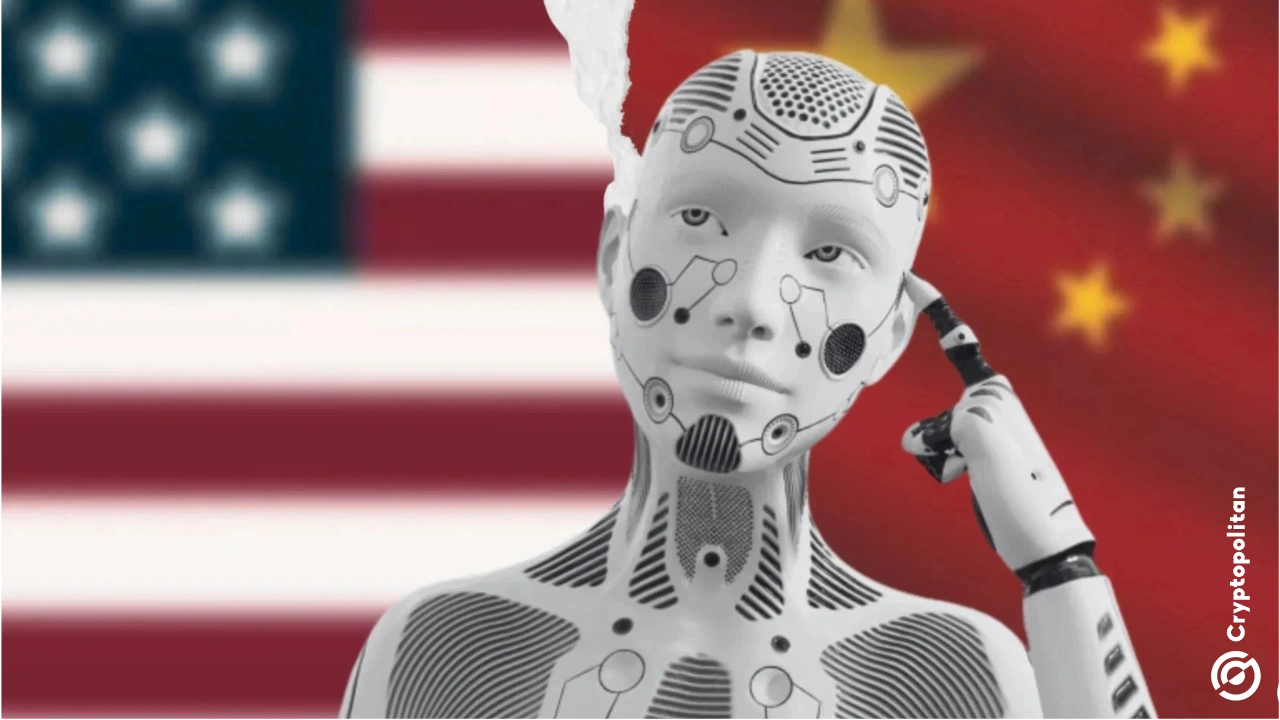
Sam OpenAI and Sundar Google are giving AI access to Pentagon-blacklisted Chinese tech giants
49 minutes ago
Tech
OpenAI floats GPT-5.6 after the Trump Admin completes security review
8 hours ago
Tech
How does Grok 4.5 compare to Anthropic’s Claude after Musk’s comments?
16 hours ago
Tech
Meta opens Muse Spark 1.1 to US developers at launch as a coding API
16 hours ago
Tech
What is going on with the global AI chip market right now?
17 hours ago
Tech
live news & hot discussions
TUNE INTO
The loudest crypto insights in real time
China AI token use explodes to 140 trillion daily with rising western share
17 hours ago
Tech
UC San Diego humanoid robots perform live surgery in world first
17 hours ago
Tech
Meta sets September start for in-house Iris AI chip production
21 hours ago
Tech
KPMG: 29% of senior business leaders struggle to understand and control AI costs
23 hours ago
Tech
OpenAI rolls out full-duplex GPT-Live voice to ChatGPT consumer users
July 9, 2026
Tech
Mistral launches Robostral Navigate, its first robotics model
July 8, 2026
Tech
Discord unbans 8,000 users after AI wrongly flagged harmless images
July 8, 2026
Tech
Musk says Grok 4.5 goes public Thursday, pitched as a cheaper Opus rival
July 8, 2026
Tech
BEST COINS'26
TON
Explore
z
ZEC
Explore
BTC
Explore
Dogecoin (DOGE)
DOGE
Explore
HYPE
Explore
China exposes security backdoor in Anthropic’s Claude Code
July 8, 2026
Tech
OpenAI’s GPT-5.6 cleared for launch after U.S. security review delayed release
July 8, 2026
Tech
Nvidia challenges Intel and AMD in $200B CPU market
July 8, 2026
Tech
Meta’s Muse Image pulls public Instagram photos into AI unless users opt out
July 8, 2026
Tech
KOR Protocol secures $7.5M from 1kx and Blockchain Capital to build AI-era IP infrastructure
July 8, 2026
Tech
B.C. attorney general moves to sue OpenAI over the Tumbler Ridge school shooting
July 7, 2026
Tech
Onsemi to sell two chip plants in $35 million cost-cutting drive
July 7, 2026
Tech
AI data centers drive Rust Belt factory power bills to record highs
July 7, 2026
Tech
Posts navigation
1
2
3
4
…
470
Next
Stay ahead in crypto
One sharp brief.
Every day.
SUBSCRIBE